About
From humble beginnings setting up Osteopathy & Physiotherapy Clinics in London, find out about our journey that has lead to us working with world class orthopaedic consultants, gaining knowledge and confidence that we want to pass on to other manual therapy and other primary healthcare practitioners.
About Us & Our Mission
Our journey into the field of musculoskeletal imaging started over 10 years ago, following a lecture given by the lead radiologists from the Royal National Orthopaedic Hospital, Dr Sajid Butt. We realised how much there had been missing from our undergraduate training as manual therapist (Osteopaths) and how much we had, until then, under appreciated the merits of good musculoskeletal imaging and reporting, particularly of the spine.
So started over a decade’s association with Dr Butt, to whom we will be eternally grateful for his expertise, patience and kindness. During this time we have spent countless hours shadowing, discussing cases and in almost daily contact with Dr Butt (and other MSK experts, including surgeons, pain consultants and other orthopaedic guru’s) gaining insights into the science and art of MRI and well as other musculoskeletal imaging (ultrasound, CT, SPECT CT, EOS etc). This has been of immense benefit to our clinical practise (spineplus.co.uk) and the patients we care for.
(In our experience) a number of features are often under appreciated by the public and by primary health care practitioners:
- Radiographers (the clinicians who select the precise areas to be scanned) do not undertake a detailed clinical examination of the patient, this is relied upon by the referrer. However, communication between the radiographer and the referrer can be lacking (see our case study on Bradley for why this is so important).
- Radiologist generate reports (MRI, X-ray etc.) but they NEVER SEE (or examine) the patient.
- There is a high degree of discrepancy (and quality) of reporting between different radiologists, see our Case Studies for examples.
- Apart from surgeons, other clinicians (GP’s, physios, chiropractors, osteopaths) who may refer for (MRI) scans, lack the training and confidence to look at the scan images and therefore rarely question radiologist or surgeon reports.
- Due to the above the potential for misunderstanding, miscommunication, misdiagnosis (false positives and false negatives) is high (we see this on a weekly basis in our clinics).
- With their expertise in face to face consultation and clinical examination, manual therapists in particular have a unique role to play for correlating clinical findings with musculoskeletal imaging. Not only will this serve the public by helping to spot mis-reporting but it will also help with patient management within clinical practice.
Go2 Imaging’s mission is to redress the imbalances outlined above and pass on the knowledge and confidence we have gained to other manual therapists and allied health professionals.
FREQUENTLY ASKED QUESTIONS
Why should I be interested in spinal imaging - studies show that just as many (if not more) people who have never had any significant back pain (or sciatica) have disc and or facet joint pathology, as those that do have pain.
The above study also showed that certain structural changes including disc herniation or radial fissures are more common in those with back pain. A study a few years after Boos et al, showed that the risk of severe back pain increased by 120% – 460% if MRI scans revealed evidence of structural changes such as spondylosis and annular tears, especially when those changes are associated with exposure to mechanical load (Videman, Leppavuori, Kaprio et al, 1998, Iatrogenice polymorphisms o the vitamin D receptor gene associated with intervertebtral disc degeneration, Spine 23 2477-85).
Regardless of this debate however, the fact is that that primary health care clinicians would be able to spot a lot more missed (serious) pathology (i.e. tumours) if they were more familiar and confident with looking at spinal MRI images. See our Case Studies for just some of the examples of this we’ve seen in clinic over the years.
Why is it though that some people escape back pain even though they have severe spinal pathology.
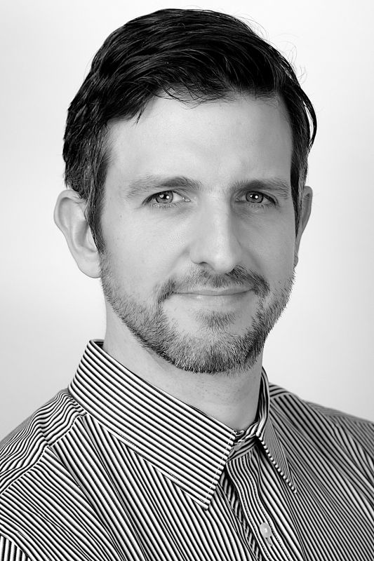
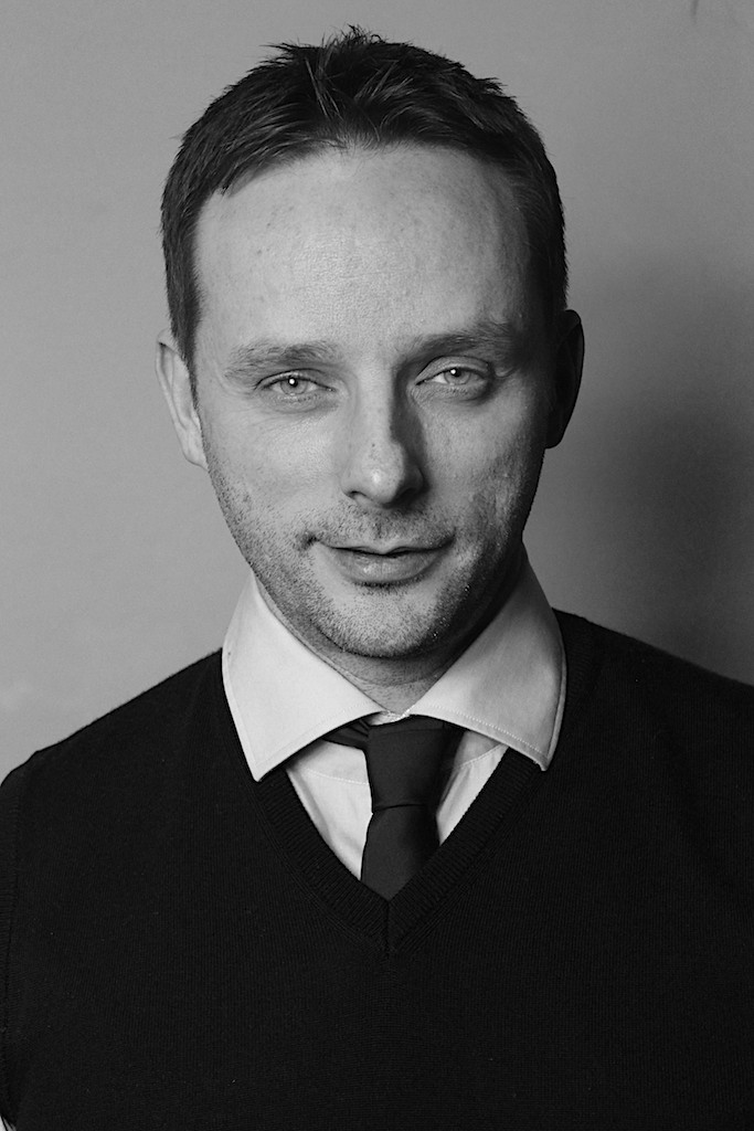
Team
We’ve spent years working with radiologists, pain consultants and surgeons. Learn with us and learn how to speak their language, engage with specialists and promote referral pathways.

Robert Shanks
Director

Darren Chandler
Director
GET NOTIFIED
Join our mailing list, so you can be notified of our training dates, webinars, guest speakers and receive FREE tips and advice.
Reach Us
Use the form opposite to drop us a message. We’ll get back to you just as soon as we can.
office@go2imaging.com
